
5 Problems With Pet monitor and How to Combat Them
Multi-parameter monitoring provides essential information for clinical diagnostic monitoring in small animal medicine. It is an important device for monitoring the vital signs of small animals
by detecting in real-time essential parameters such as ECG signal, heart rate, oxygen saturation, blood pressure, respiratory rate, body temperature, and end-expiratory carbon dioxide.
Proficiency in the operation of the ECG monitor is a skill that every veterinarian should have.
What are the key operational points that should not be overlooked for training the use of the pet monitor?
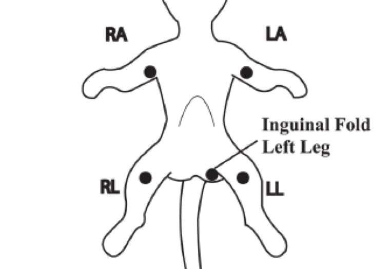
I. What is the difference between a 5-lead and a 3-lead ECG lead?
3 lead ECG leads can only be used to obtain I, II, and III show ECGs, whereas 5 lead ECG leads can be used to obtain I, II, III, AVR, AVF, AVL, and V lead ECGs.
To facilitate quick connection, color coding is used to allow quick clamping to the appropriate clip. 3 lead ECG leads are color-coded (American Standard) white (RA), black (LA), and red (LL),
and 5 lead ECG leads are color-coded white (RA), black (LA), red (LL), green (RL), and brown (V).
The position of the ECG clips is not the same for leads of the same color in both sizes, and it is more reliable to use the abbreviations RA, LA, RL, LL, and V to determine the position than to
memorize the colors. In the dog, for example, the electrodes of the standard lead, the unipolar pressurized limb lead, should be placed above the hawk's beak and on the patellar ligament in a
similar way to the human.
II. Can the oximetry clip and blood pressure cuff be placed on the same limb?
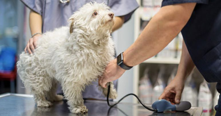
Blood pressure measurement can block arterial blood flow, resulting in inaccurate oxygen saturation levels during blood pressure measurement, so it is not clinically recommended that
the oxygen saturation clip and blood pressure cuff be
placed on the same limb.
III. Why is the II-lead ECG waveform often used?
The waveform is most obvious on an II-lead ECG. When recording an ECG at the bedside, in addition to the usual 12-lead simultaneous recording, a separate long strip of II-lead recording is
also required, mainly to facilitate comparative analysis
and to facilitate the analysis of various arrhythmias.

IV. The monitor I bought does not have an end-expiratory CO2 function and there is no module to monitor respiration, how is respiration monitored?
The respiration on the monitor relies on the electrical clips of the ECG to sense changes in the impedance of the animal's thorax and to display the respiratory waveform and data. As the lower
left and upper right clips are the respiratory
sensing positions, it is important where they are clipped.
The 2 clips should be placed diagonally as far as possible to obtain the best respiratory waveform.

V. Why are there often large deviations in the first measurement when a new sick animal has its blood pressure measured with a cardiac monitor?
Many ECG monitors have a memory storage function, so if a hypertensive animal was last used and the measurement was not cleared to zero after use, then the next time another animal with
normal blood pressure uses the monitor for the
the first time the results are likely to be high, as the monitor is automatically calibrated, the second measurement will usually be close to the animal's state.
Note: The monitors are used to help animal doctors do their job better. Each instrument has detailed instructions for use, so please read them carefully before using them correctly.
We're always here to help! Contact us with any questions or concerns.
View Product
Hyperbaric Therapy: One Real Animal's Success Story
New Treatment for Cat-borne abdomen You Should Know About
Related Article


