
Intraocular Pressure (IOP) Measurements in Animals
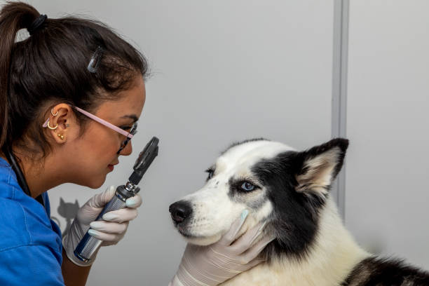 Intraocular pressure (IOP) measurement is a crucial aspect of veterinary ophthalmology, allowing veterinarians to assess the health of an animal's eyes and detect ocular abnormalities. Proper interpretation of IOP values is essential for accurate diagnosis and effective management of various eye conditions. In this article, we will explore the significance of IOP measurement, methods for obtaining accurate readings, factors influencing IOP values, and the importance of interpreting these values correctly in animals. I. Importance of Intraocular Pressure Measurement Intraocular pressure plays a vital role in maintaining the shape and health of the eye. It is the pressure exerted by the fluid inside the eye (aqueous humor) against the walls of the eyeball. Normal IOP is essential for proper ocular function and vision. Abnormalities in IOP levels can indicate underlying eye diseases such as glaucoma, uveitis, or ocular trauma. Therefore, measuring IOP is a fundamental part of ocular examination in veterinary medicine. II. Methods for Obtaining Accurate IOP Readings Several techniques are available for measuring IOP in animals, including applanation tonometry, indentation tonometry, and rebound tonometry. Applanation tonometry, such as the Goldmann tonometer, is considered the gold standard for accurate IOP measurement in both humans and animals. The procedure involves applying a precise amount of pressure to the cornea and measuring the force required to flatten a specific area. Proper preparation of the animal, adequate sedation if necessary, and instrument calibration are essential for obtaining reliable IOP readings. III. Factors Affecting Intraocular Pressure Measurement Various factors can influence IOP values in animals, including species variability, age, breed, body size, ocular pathologies, and systemic diseases. Normal IOP ranges may vary between different species and breeds, making it crucial to consider these factors when interpreting IOP values. Additionally, certain conditions such as corneal edema, uveitis, or systemic hypertension can affect IOP readings and require careful evaluation. IV. Interpreting Intraocular Pressure Values Interpreting IOP values involves comparing the measured pressure to established normal ranges for the species and breed of the animal. Normal IOP values may range from 10 to 20 mmHg in dogs and cats, but there can be variations depending on factors such as age and breed. Elevated IOP (hypertony) may indicate conditions like glaucoma, while decreased IOP (hypotony) may be associated with uveitis or trauma. Serial IOP measurements over time are often necessary to monitor changes and assess treatment response accurately. V. Clinical Considerations and Differential Diagnoses In clinical practice, veterinarians must consider differential diagnoses based on IOP values and other clinical findings. Elevated IOP may prompt further evaluation for conditions such as primary or secondary glaucoma, while decreased IOP may indicate uveitis, lens luxation, or systemic hypotension. Integration of IOP measurements into comprehensive ophthalmic examinations, including slit-lamp biomicroscopy, fundoscopy, and ocular ultrasonography, can aid in the diagnosis and management of ocular diseases. VI. Case Studies and Examples Case studies provide valuable insights into the practical application of IOP measurement in veterinary ophthalmology. For example, a case of elevated IOP in a canine patient with acute onset of ocular pain and corneal edema may raise suspicion for primary angle-closure glaucoma. Conversely, chronically low IOP in a feline patient with recurrent episodes of anterior uveitis may indicate underlying systemic diseases such as feline infectious peritonitis (FIP) or lymphoma. VII. Management Strategies Based on IOP Findings Management strategies for ocular conditions are often tailored to the underlying cause and severity of the disease. In cases of elevated IOP, treatment may involve medical therapy, such as topical or systemic medications, laser therapy, or surgical intervention (e.g., trabeculectomy). Conversely, managing hypotony may require addressing the underlying inflammation or trauma, providing supportive care, and monitoring for complications such as retinal detachment. In conclusion, understanding and interpreting intraocular pressure measurements are essential skills for veterinary ophthalmologists and general practitioners. Accurate assessment of IOP values allows for early detection, diagnosis, and appropriate management of ocular conditions in animals. By integrating IOP measurement into routine ocular examinations and clinical decision-making, veterinarians can optimize patient care and improve visual outcomes for their furry patients. |
Navigating Pet Hospital Visits: Essential vs. Optional Examinations
Winning Tips for Dog Joint Care
Related Article


