
Veterinary Imaging Room Solution
Build the right veterinary imaging capability for your clinic or animal hospital—from a new X-ray room or DR upgrade to ultrasound or a combined imaging setup
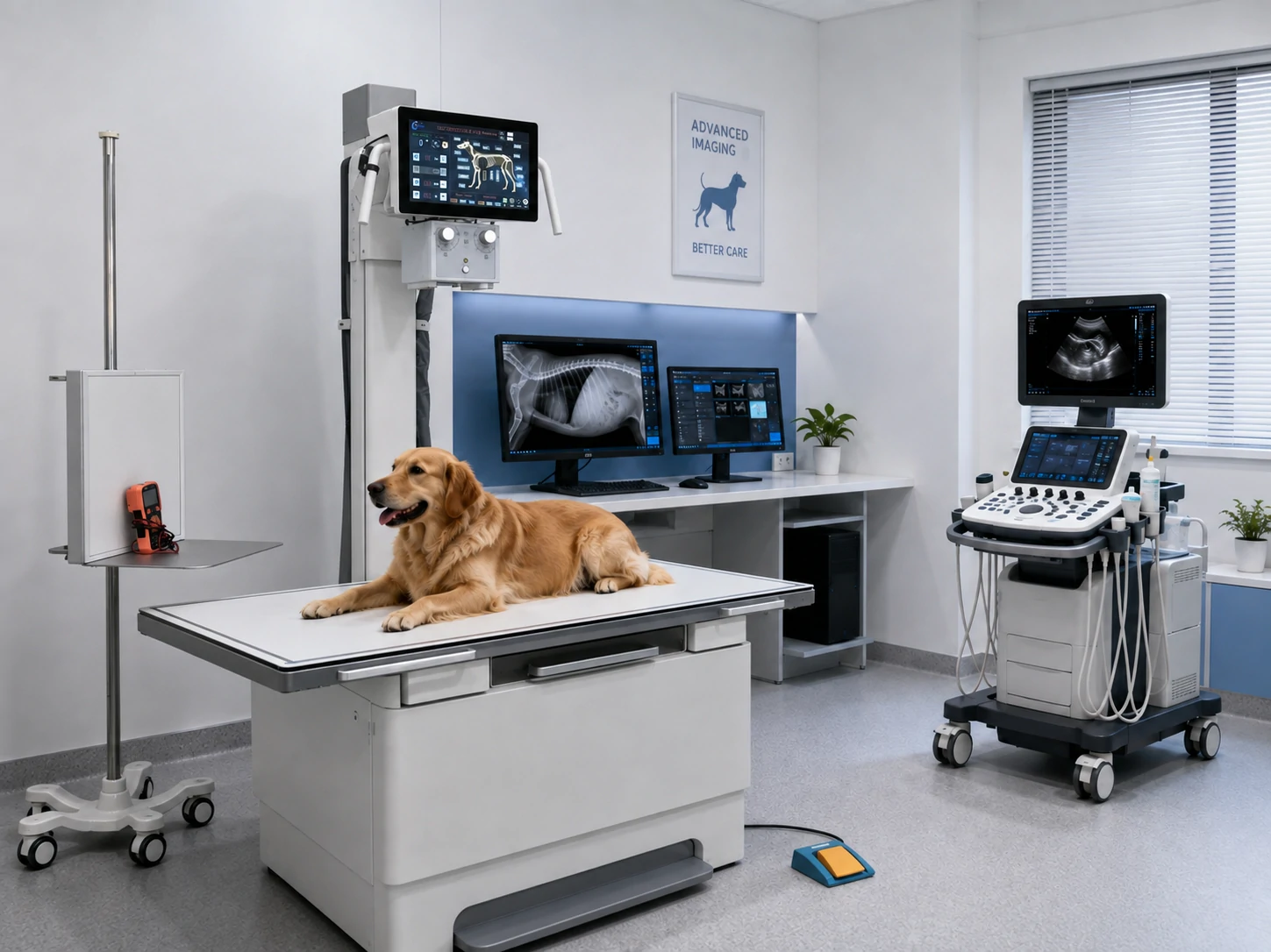
Choose the Right Veterinary Imaging Modality
Build the right veterinary imaging capability for your clinic or animal hospital—from a new X-ray room or DR upgrade to ultrasound or a combined imaging setup
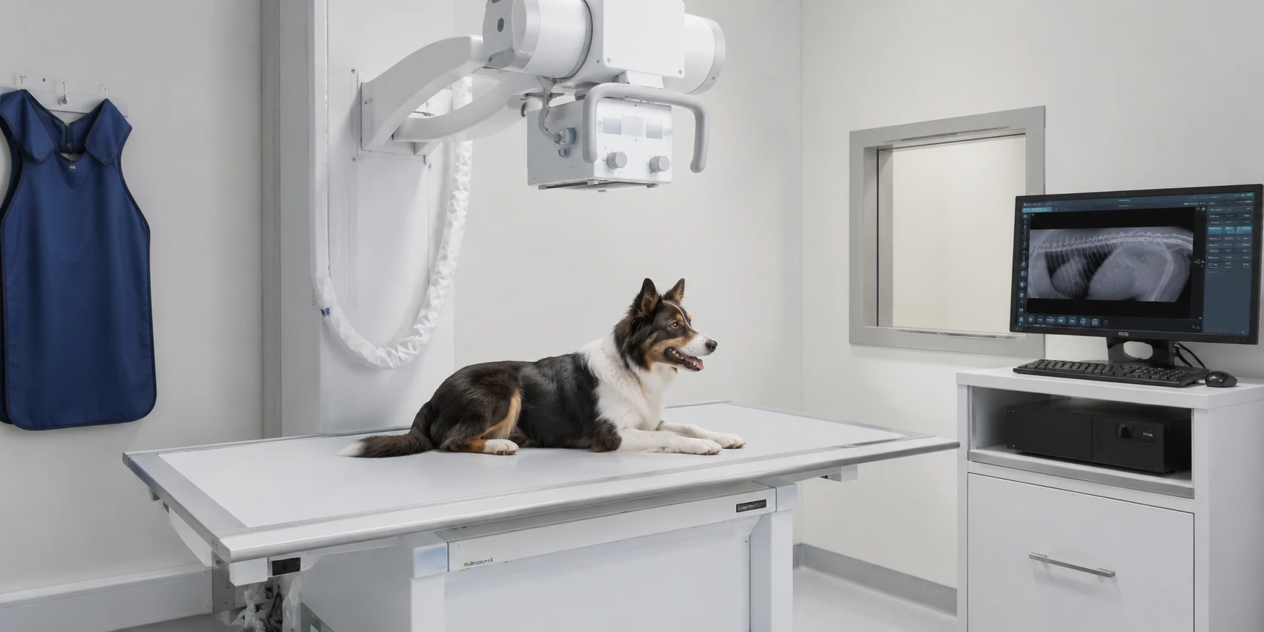
VETERINARY X-RAY
Better for radiography-focused needs
- Skeletal and trauma overview
- Thoracic examinations
- Routine radiography
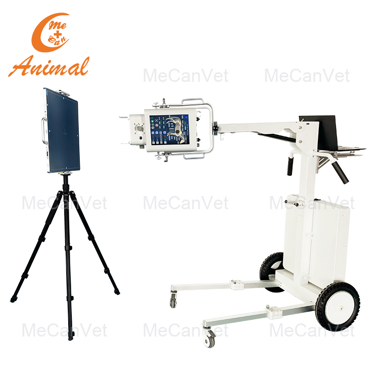
Typical direction: Fixed, mobile, or portable X-ray with DR, workstation, and positioning support.
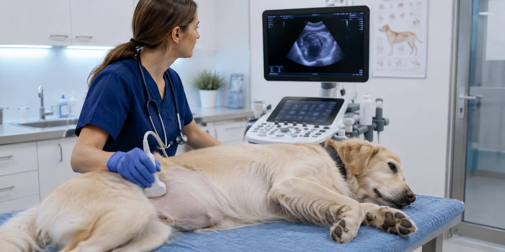
VETERINARY ULTRASOUND
Better for soft-tissue imaging
- Abdominal and organ evaluation
- Reproductive applications
- Selected advanced applications if supported
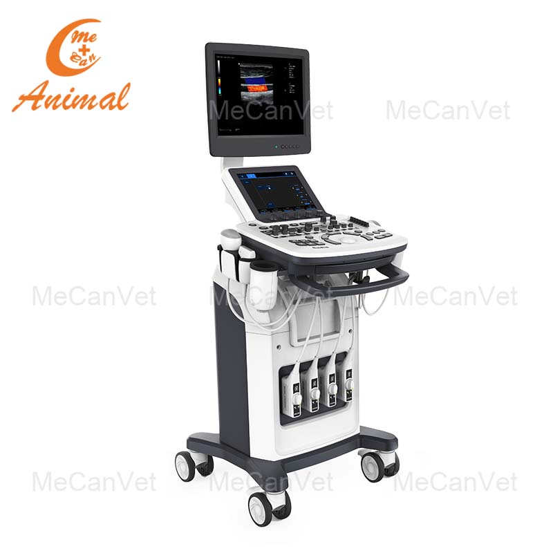
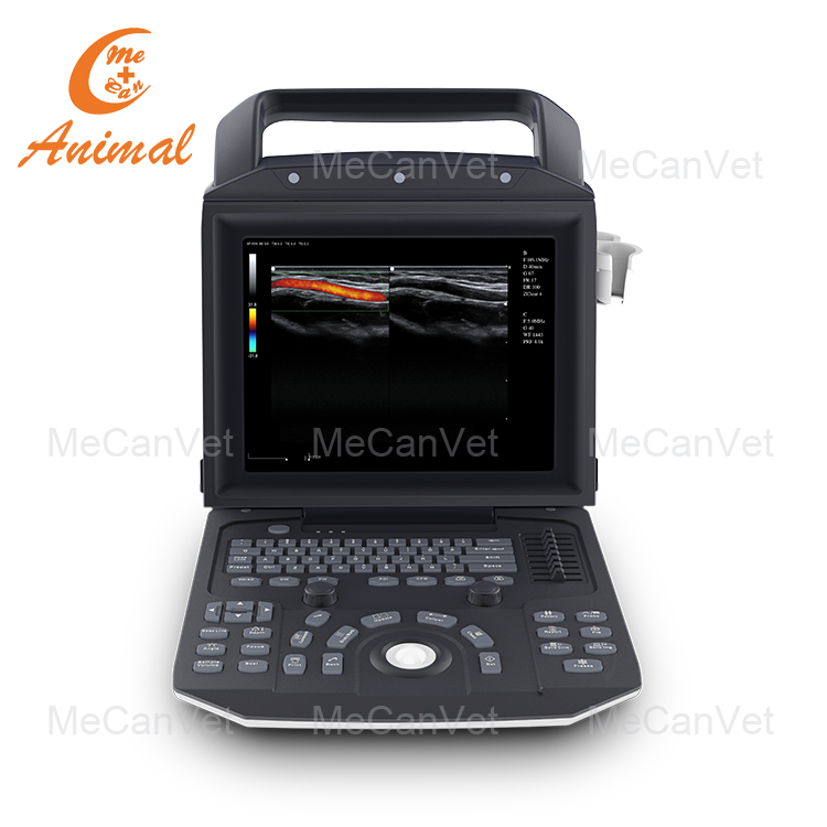
Typical direction: Portable or cart-based ultrasound matched to supported probes and animal size.
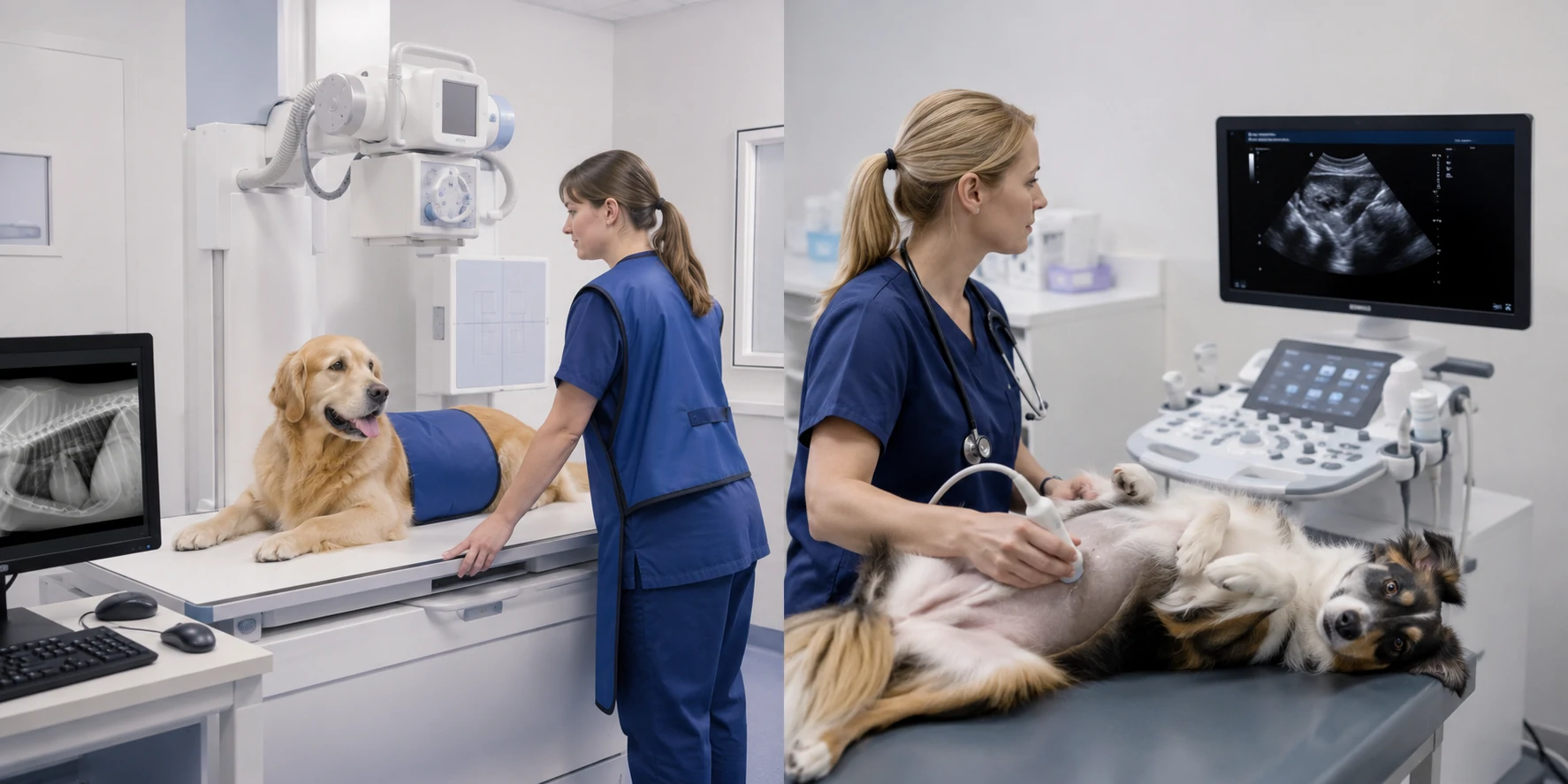
X-RAY + ULTRASOUND
Broader in-house imaging capability
- Complementary modalities
- Shared or separate workflow
- Core, optional or phased purchasing
No fixed package.: Room arrangement depends on workload, space and facility workflow.
What You Will Receive
Veterinary X-ray Room Setup — Philippines
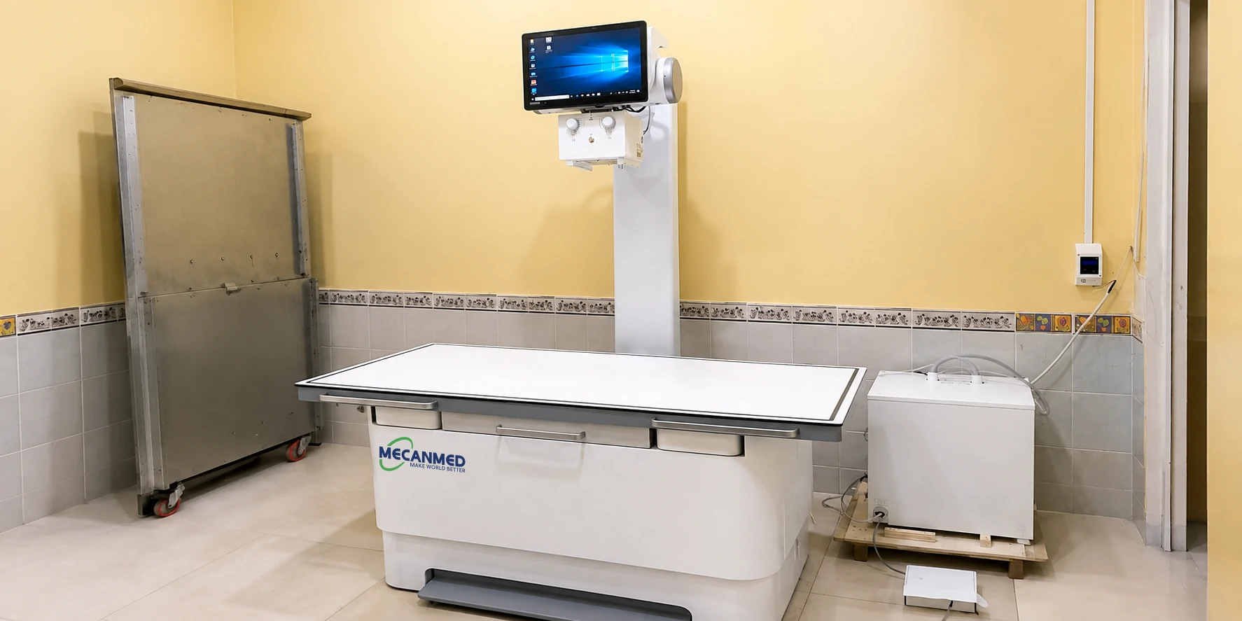
REAL PROJECT PROOF
In-house digital radiography capability
Project Need
Add in-house digital radiography.
Matched Configuration
Veterinary X-ray system, imaging table and acquisition workstation.
MeCan Vet Support
Equipment supply, setup communication and on-site user training for this project.
Build Your Veterinary X-ray Configuration
A veterinary X-ray setup should be selected around examination needs, imaging workload, positioning and room conditions rather than a fixed package.
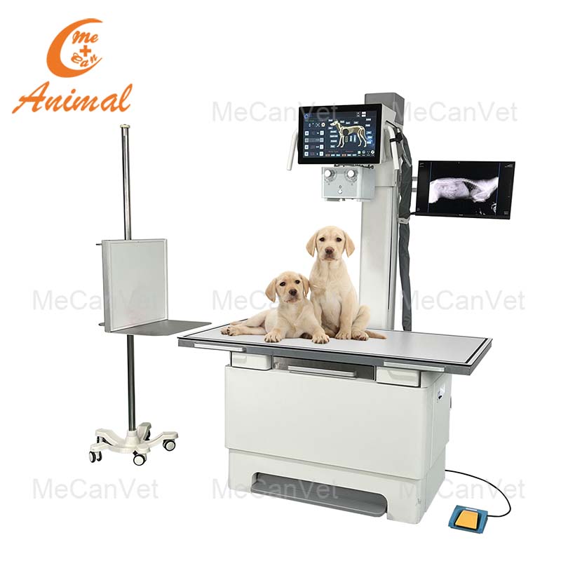
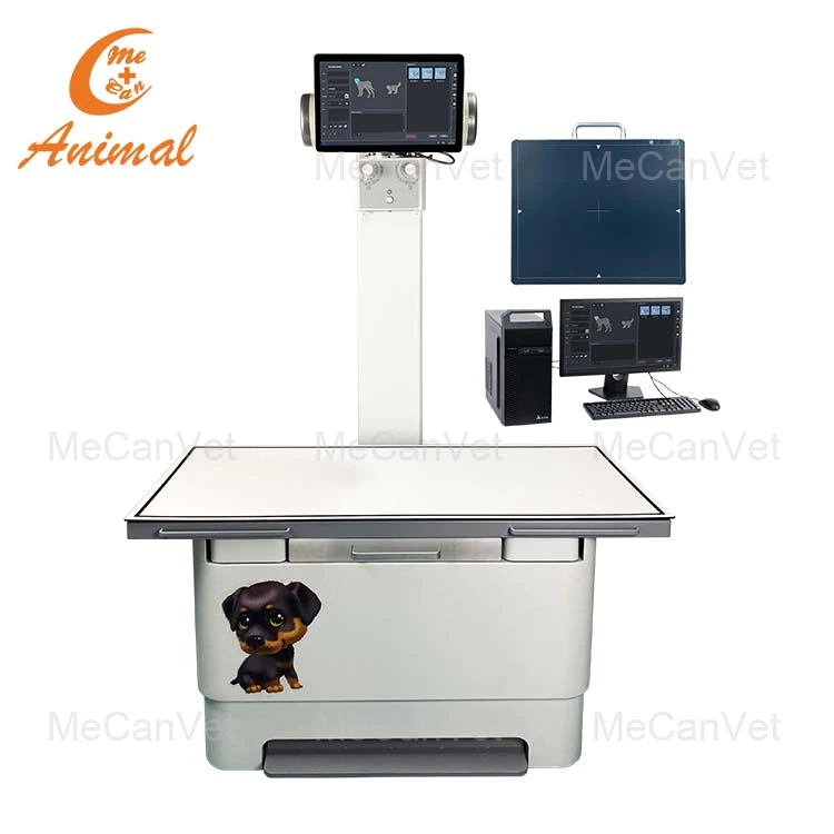
Fixed X-ray
Consider for dedicated rooms with a stable and potentially higher-frequency workflow.
Mobile X-ray
Consider when equipment movement inside the facility is important.
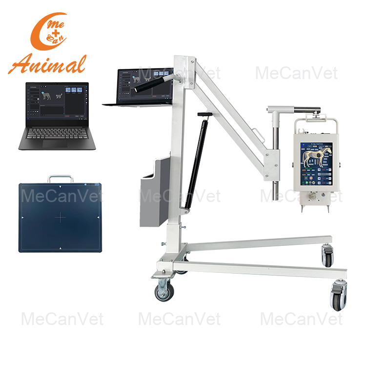
Portable X-ray
Consider where space, flexible positioning or mobile use is important.
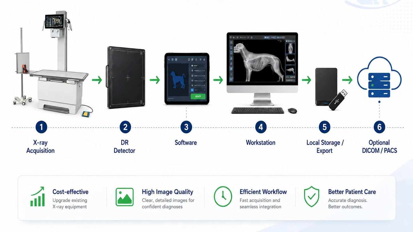
DIGITAL RADIOGRAPHY WORKFLOW
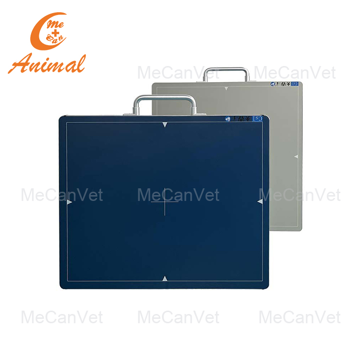
DR is more than the X-ray generator alone
X-ray System
DR Detector
Acquisition Software
Workstation
Review / Storage
For upgrade projects, review generator, exposure interface, detector size,
workstation and software together before detector selection.
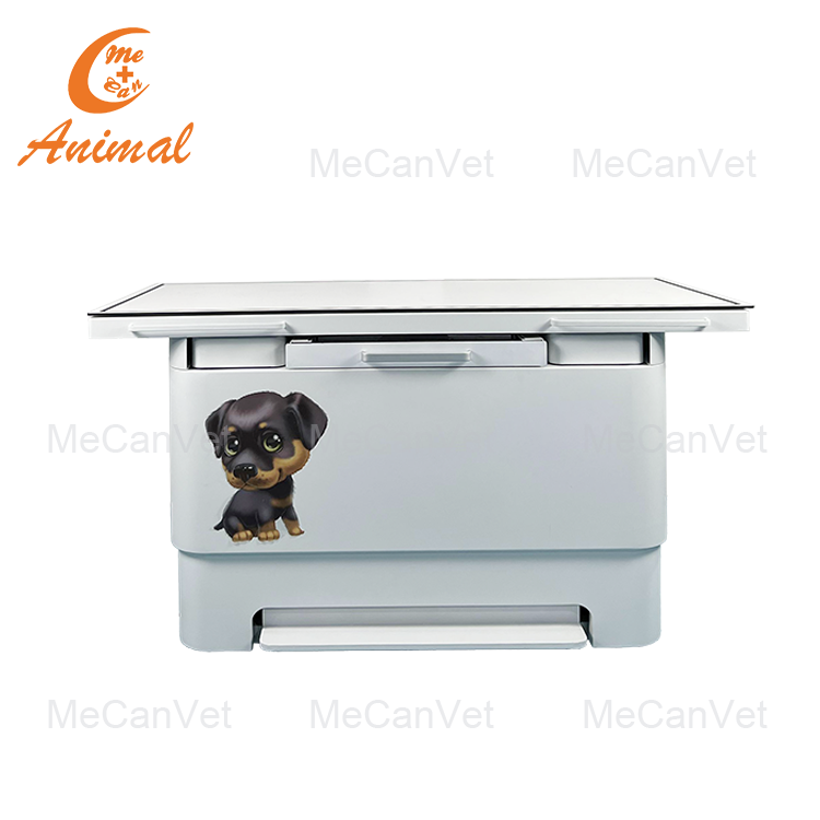
Veterinary Imaging Table & Positioning Support
Animal size and weight range
Table or standing workflow
Detector placement and support
Positioning aids
Animal entry and transfer path
Consider when equipment movement inside the facility is important.
Choose the Right Veterinary Ultrasound Setup
Select veterinary ultrasound according to planned applications, animal size, mobility and supported probes—not by feature count alone.
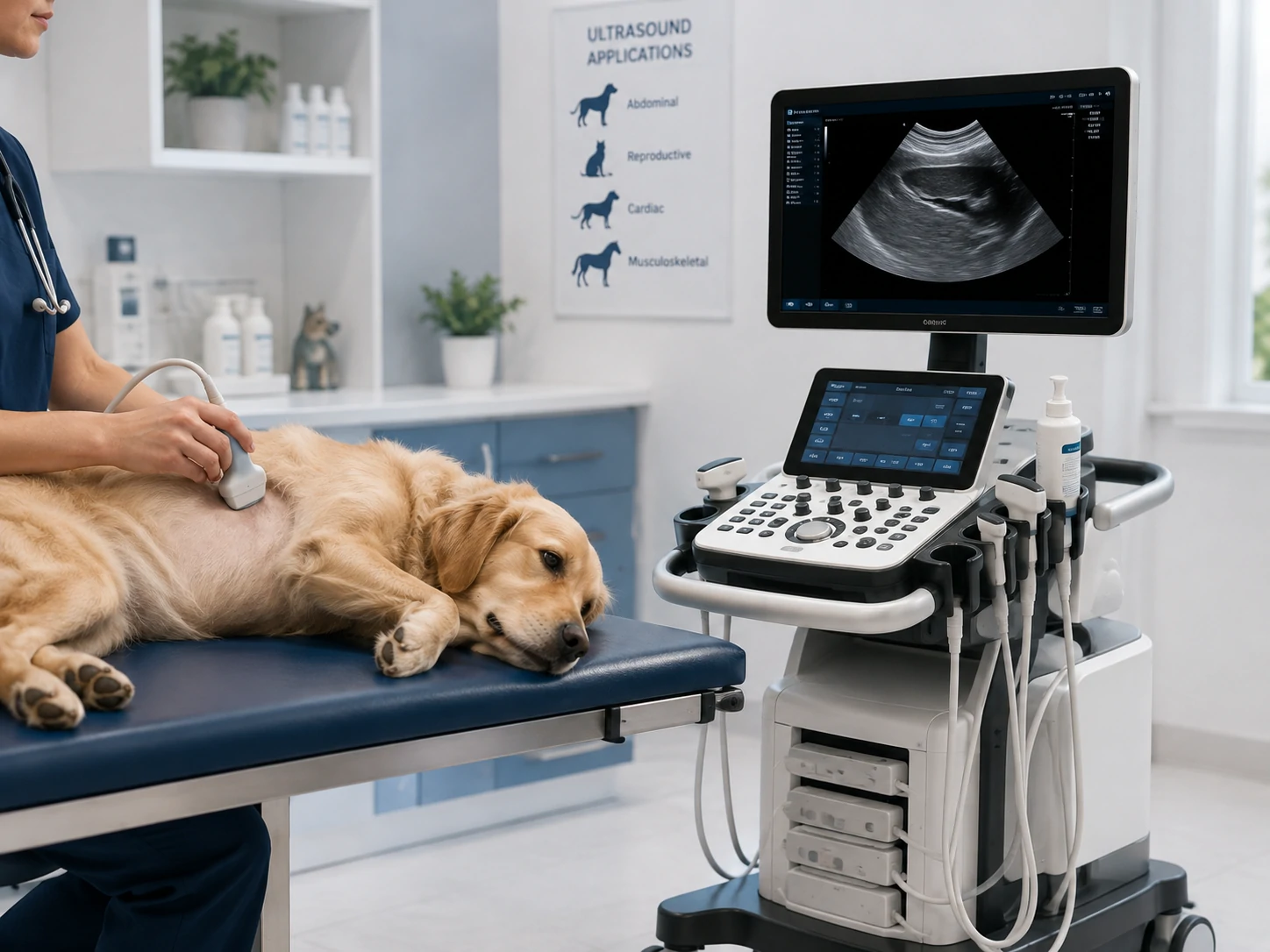
Portable Ultrasound
Suitable for limited space, room-to-room use or mobile and field applications.
Cart-Based Ultrasound
Suitable for dedicated examination areas, frequent ultrasound use or multi-probe workflows where supported.
Applications & Probe Selection
Match the ultrasound system and supported probes to planned examinations and animal size.
Abdominal
Reproductive
Color Doppler — if required
Cardiac — if supported
Probe availability and advanced applications depend on the selected ultrasound system.
Veterinary Positioning & Imaging Workflow
Animal movement, positioning and detector access should be considered before the final equipment configuration is confirmed.
1. Preparation
Exam plan & animal condition
2. Transfer & Positioning
Table, standing or mobile workflow
3. Image Acquisition
Stable positioning & detector access
4. Review
Workstation or ultrasound display
5. Storage / Clinical Use
Local storage, export or integration
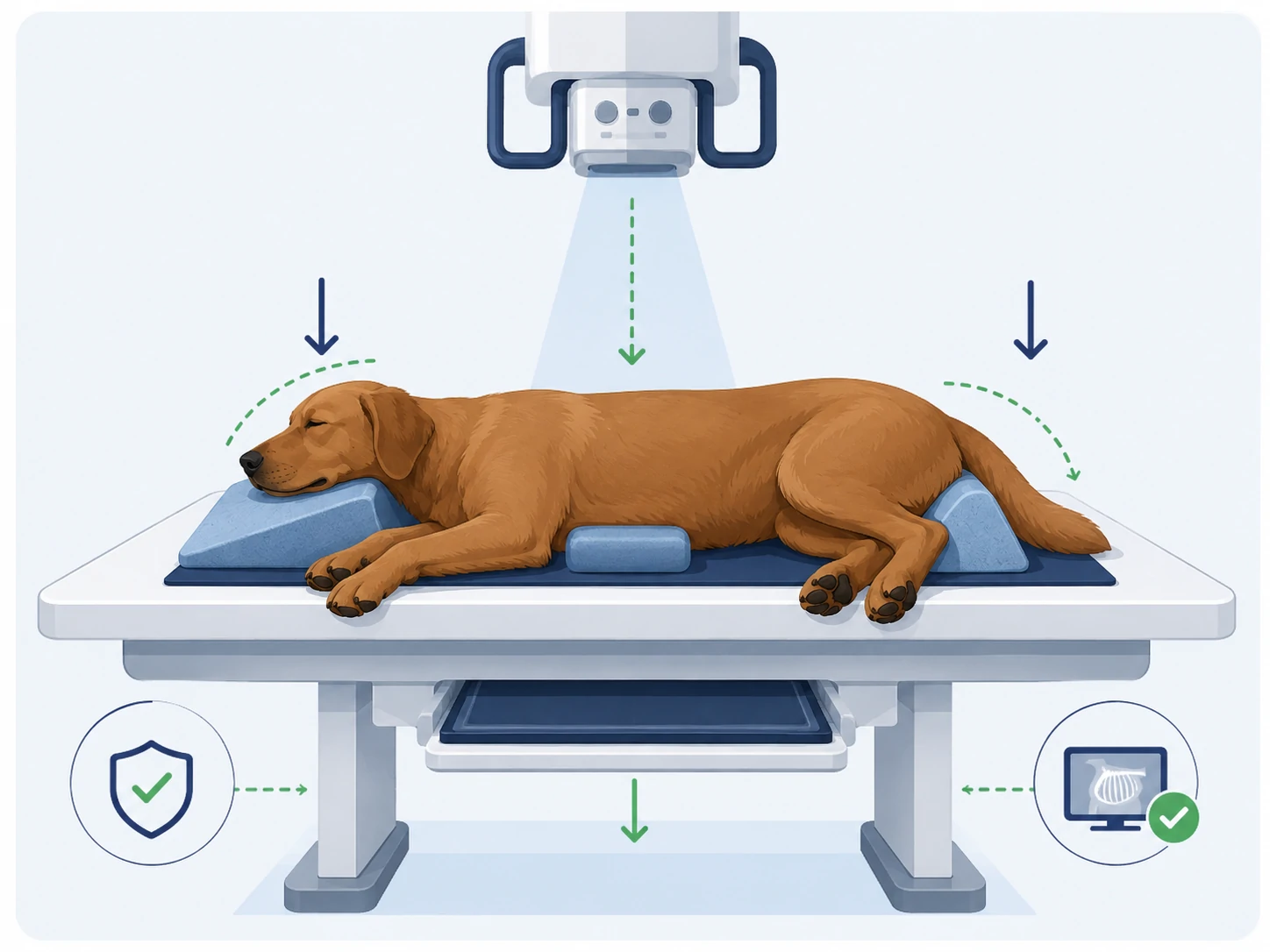
Stable Positioning
Support consistent positioning and reduce avoidable motion.
Positioning Aids
Use appropriate supports to reduce unnecessary manual restraint during exposure.
Radiation-Safe Workflow
Staff protection and exposure procedures must follow applicable local requirements.
Animal handling, restraint and sedation decisions remain the veterinary team’s responsibility and should be determined case by case.
Imaging Room Layout & Radiation Considerations
Room planning should consider equipment footprint, animal movement, operator workflow, service access and local radiation requirements.
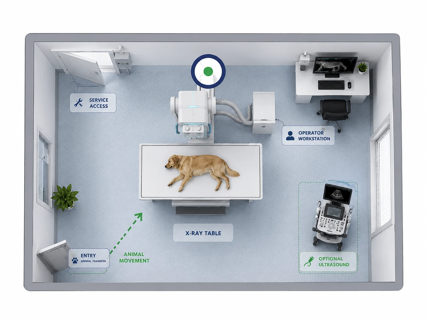
Shared or Separate Ultrasound Area?
It depends on available space, workload, radiation workflow and facility scale. A smaller clinic may use a flexible arrangement, while a higher-volume animal hospital may benefit from separate or dedicated imaging spaces.
Radiation Considerations
Final shielding design, calculations, construction, permits and approvals should be handled by qualified local parties.
DR Upgrade & Digital Imaging Workflow
Existing veterinary X-ray equipment may be suitable for DR conversion, but compatibility should be reviewed before detector selection.
Already Have an X-ray System?
Share available information for an initial review:
Key Configuration Conditions
These details help screen the right imaging direction before the final list and quotation.
Animal & Examination
Species, weight range, body areas and planned examinations.
Imaging Workload
Estimated imaging frequency, number of users and typical workflow.
Room & Utilities
Available dimensions, access, power, grounding and network conditions.
Existing Equipment
Generator, detector, workstation and software information for upgrade projects.
Positioning & Mobility
Animal transfer, table or standing workflow, detector handling and mobile use.
Local Radiation Requirements
Protection and approval requirements confirmed with qualified local professionals.
Have partial information only?
That is enough to start an initial configuration review.
Recommended Veterinary
Imaging Equipment




Get Your Veterinary Imaging Configuration
Tell us your animal types, planned examinations and available room or existing equipment information. If you are not sure whether you need X-ray, ultrasound or both, start with your diagnostic needs.
New imaging room
Existing X-ray → DR upgrade
Ultrasound expansion
Combined X-ray + ultrasound